Файл:Phytomyxea collage.jpg
Перейти к навигации
Перейти к поиску

Размер этого предпросмотра: 501 × 600 пкс. Другие разрешения: 200 × 240 пкс | 600 × 718 пкс.
Исходный файл (600 × 718 пкс, размер файла: 201 КБ, MIME-тип: image/jpeg)
Этот файл находится на Викискладе. Сведения о нём показаны ниже.
Викисклад — централизованное хранилище для свободных файлов, используемых в проектах Викимедиа.
|
Краткое описание
| Описание |
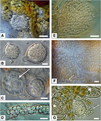
English: Morphology of resting spores from selected phytomyxids. Bar=10 μm.
|
| Дата | |
| Источник | Neuhauser S., Kirchmair M., Bulman S., Bass D. (2014). "Cross-kingdom host shifts of phytomyxid parasites". BMC Evolutionary Biology 14 (33). DOI:10.1186/1471-2148-14-33. |
| Автор | Sigrid Neuhauser, Martin Kirchmair, Simon Bulman and David Bass |
Лицензирование
Этот файл доступен по лицензии Creative Commons Attribution 2.0 Generic
|
This file was published in a BioMed Central journal. Their website states that all of its research publications is published under the license which is identical to the Creative Commons Attribution 2.0 license (some non-research articles like reviews or editorials may require a subscription.)
To the uploader: You must provide a link (URL) to the original file or journal article.
|
История файла
Нажмите на дату/время, чтобы посмотреть файл, который был загружен в тот момент.
| Дата/время | Миниатюра | Размеры | Участник | Примечание | |
|---|---|---|---|---|---|
| текущий | 13:38, 24 января 2016 | | 600 × 718 (201 КБ) | Mithril | =={{int:filedesc}}== {{Information |Description= {{en|1=Morphology of resting spores from selected phytomyxids. Bar=10 μm: '''left column''': Plasmodiophorida, '''right column''': Phagomyxida. *'''A''': ''Sorosphaerula viticola'': hollow sporosori in... |
Использование файла
Следующая страница использует этот файл:
Глобальное использование файла
Данный файл используется в следующих вики:
- Использование в ar.wikipedia.org
- Использование в arz.wikipedia.org
- Использование в en.wikipedia.org
- Использование в es.wikipedia.org
- Использование в ia.wikipedia.org
- Использование в ko.wikipedia.org
- Использование в pl.wikipedia.org
- Использование в ro.wikipedia.org
- Использование в tr.wikipedia.org
- Использование в www.wikidata.org